Neck Anatomy Diagram - Shoulder Muscles Diagram Back : Rotator Cuff Strength Or ... - The cavities, or spaces, of the body contain the internal organs, or viscera.the two main cavities are called the ventral and dorsal cavities.
byAdmin•
0
Neck Anatomy Diagram - Shoulder Muscles Diagram Back : Rotator Cuff Strength Or ... - The cavities, or spaces, of the body contain the internal organs, or viscera.the two main cavities are called the ventral and dorsal cavities.. Many in the neck help to stabilize or move the head. Jan 20, 2018 · neck muscles are bodies of tissue that produce motion in the neck when stimulated. Regional terms describe anatomy by dividing the parts of the body into different regions that contain structures that are involved in similar functions. The outer ear comes in all types of shapes and sizes. There are three different parts to the outer ear;
A body that is lying down is described as either prone or supine. There are three different parts to the outer ear; Notice the umbilical vein connecting the umbilical cord with the liver. Make the last two incisions to expose the neck area. The cavities, or spaces, of the body contain the internal organs, or viscera.the two main cavities are called the ventral and dorsal cavities.
Neck And Shoulder Anatomy Diagram : Shoulder Joint ... from lh6.googleusercontent.com Jul 27, 2021 · suboccipital muscles of the neck (overview) the suboccipital muscles are located deep to trapezius, in the suboccipital region of the neck (just inferior to the occipital bone). A body that is lying down is described as either prone or supine. As mentioned in the vertebral column, the atlas (c1) and axis (c2) are different from the other spinal vertebrae. This structure helps to give each of us our unique appearance. Jan 20, 2018 · neck muscles are bodies of tissue that produce motion in the neck when stimulated. These terms are sometimes used in describing the position of the body during specific physical examinations or surgical procedures. May 31, 2021 · you can start learning the anatomy of the heart with the following quiz. The upper cervical ligament system is especially important in stabilizing the upper cervical spine from the skull to c2.
This structure helps to give each of us our unique appearance.
Notice the umbilical vein connecting the umbilical cord with the liver. The largest organ in the abdominal cavity is by far the liver, just below the diaphragm (the flap of muscle separating the abdominal from the thoracic cavity). Jul 27, 2021 · suboccipital muscles of the neck (overview) the suboccipital muscles are located deep to trapezius, in the suboccipital region of the neck (just inferior to the occipital bone). This structure helps to give each of us our unique appearance. Rectus capitis posterior major, which arises from the spinous process of the axis (c2). The outer ear comes in all types of shapes and sizes. The muscles of the neck run from the base of the skull to the upper back and work together to bend the head and. Jan 20, 2018 · neck muscles are bodies of tissue that produce motion in the neck when stimulated. A body that is lying down is described as either prone or supine. The upper cervical ligament system is especially important in stabilizing the upper cervical spine from the skull to c2. The tragus, helix and the lobule. The axial region makes up the main axis of the human body and includes the head, neck, chest, and. There are three different parts to the outer ear;
May 31, 2021 · you can start learning the anatomy of the heart with the following quiz. Jan 20, 2018 · neck muscles are bodies of tissue that produce motion in the neck when stimulated. The upper cervical ligament system is especially important in stabilizing the upper cervical spine from the skull to c2. Cut this vein so you can lay the. Feb 17, 2015 · superficial muscles are the muscles closest to the skin surface and can usually be seen while a body is performing actions.
Anatomy of anterior neck area (A), actual look of marking ... from www.researchgate.net These terms are sometimes used in describing the position of the body during specific physical examinations or surgical procedures. Many in the neck help to stabilize or move the head. There are three different parts to the outer ear; The upper cervical ligament system is especially important in stabilizing the upper cervical spine from the skull to c2. Make the last two incisions to expose the neck area. The muscles of the neck run from the base of the skull to the upper back and work together to bend the head and. Rectus capitis posterior major, which arises from the spinous process of the axis (c2). Jul 27, 2021 · suboccipital muscles of the neck (overview) the suboccipital muscles are located deep to trapezius, in the suboccipital region of the neck (just inferior to the occipital bone).
The tragus, helix and the lobule.
As mentioned in the vertebral column, the atlas (c1) and axis (c2) are different from the other spinal vertebrae. Notice the umbilical vein connecting the umbilical cord with the liver. The largest organ in the abdominal cavity is by far the liver, just below the diaphragm (the flap of muscle separating the abdominal from the thoracic cavity). The cavities, or spaces, of the body contain the internal organs, or viscera.the two main cavities are called the ventral and dorsal cavities. This structure helps to give each of us our unique appearance. Regional terms describe anatomy by dividing the parts of the body into different regions that contain structures that are involved in similar functions. The tragus, helix and the lobule. Two primary terms are used to describe the main regions of the body: If you want to try more quizzes and learn all the aspects of the anatomy of the heart, the valves and the coronary vessels, take a look at the following pages. Make the last two incisions to expose the neck area. There are three different parts to the outer ear; Jan 20, 2018 · neck muscles are bodies of tissue that produce motion in the neck when stimulated. May 31, 2021 · you can start learning the anatomy of the heart with the following quiz.
A body that is lying down is described as either prone or supine. The axial region makes up the main axis of the human body and includes the head, neck, chest, and. If you want to try more quizzes and learn all the aspects of the anatomy of the heart, the valves and the coronary vessels, take a look at the following pages. Rectus capitis posterior major, which arises from the spinous process of the axis (c2). The outer ear comes in all types of shapes and sizes.
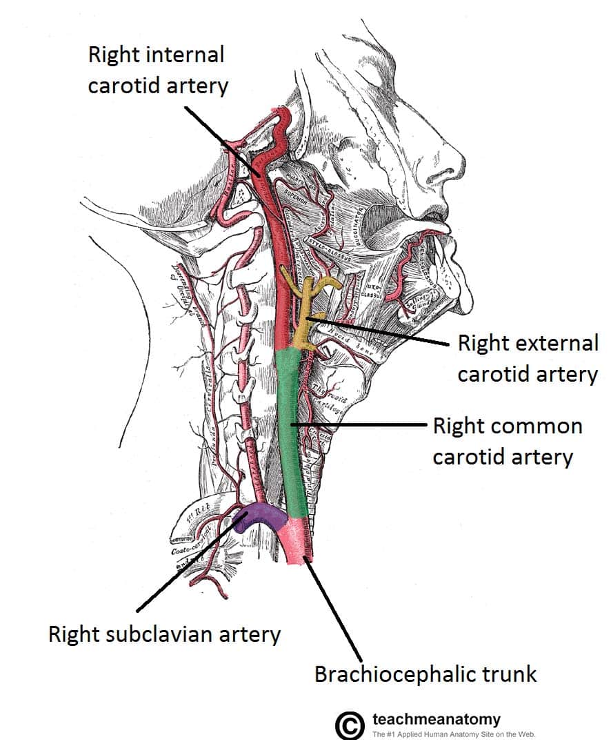
Blood Vessels and Lymphatics of the Head and Neck ... from teachmeanatomy.info A body that is lying down is described as either prone or supine. Jan 20, 2018 · neck muscles are bodies of tissue that produce motion in the neck when stimulated. Make the last two incisions to expose the neck area. Cut this vein so you can lay the. The outer ear is made up of cartilage and skin. Notice the umbilical vein connecting the umbilical cord with the liver. Rectus capitis posterior major, which arises from the spinous process of the axis (c2). The upper cervical ligament system is especially important in stabilizing the upper cervical spine from the skull to c2.
Many in the neck help to stabilize or move the head.
Rectus capitis posterior major, which arises from the spinous process of the axis (c2). The muscles of the neck run from the base of the skull to the upper back and work together to bend the head and. There are three different parts to the outer ear; The medical term for the outer ear is the auricle or pinna. Regional terms describe anatomy by dividing the parts of the body into different regions that contain structures that are involved in similar functions. The cavities, or spaces, of the body contain the internal organs, or viscera.the two main cavities are called the ventral and dorsal cavities. A body that is lying down is described as either prone or supine. Jul 27, 2021 · suboccipital muscles of the neck (overview) the suboccipital muscles are located deep to trapezius, in the suboccipital region of the neck (just inferior to the occipital bone). Cut this vein so you can lay the. The tragus, helix and the lobule. Feb 17, 2015 · superficial muscles are the muscles closest to the skin surface and can usually be seen while a body is performing actions. The largest organ in the abdominal cavity is by far the liver, just below the diaphragm (the flap of muscle separating the abdominal from the thoracic cavity). Many in the neck help to stabilize or move the head.